
English / Español
NOT A SCIENTIST?
A lay summary of our research
From the moment of our conception and throughout our growth, our cells undergo millions of cycles of division to orchestrate the formation of our bodies, including our bones, heart, bran and more. Furthermore, within our own mature bodies, approximately two trillion cells divide daily to renew the cells that make up our skin, blood, intestine, etc.
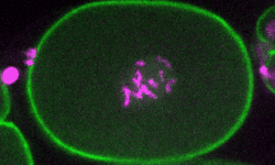
A movie of a C. elegans embryo as it undergoes cell division.
Throughout these intricate processes, it is crucial that cells to divide at the right time and at the right place. To ensure this precision, cells receive a multitude of “signals” that either prompt them to divide or instruct them to pause if they are not ready. These signals may originate externally, such as from neighboring cells; or internally, such as assessing their nutritional status. Following on the assessment of these signals, a cell can decide to either divide or pause. Errors in this decision process can have catastrophic consequences. On one hand, uncontrolled cell division can lead to cancer, while, on the other hand, failure to divide is associated with several developmental abnormalities.
In our laboratory, we explore how cells integrate diverse and sometimes conflicting signals during development to either divide, pause or become differentiated. For this, we use a combination of technologies that include genome editing, genetic screens, live-imaging microscopy, biochemistry and proteomic analyses.

A plate with C. elegans under a fluorescence stereo microscope
Our primary system for many of these studies is the microscopic worm C. elegans, which lives in the soil and compost, but can be easily cultivated in the laboratory.
Why do we use worms for studying cell division? Surprisingly, worms and humans are more similar than anticipated. In fact, we share the same genes that participate in cell division. However, unlike humans, we can easily manipulate worms, such as by editing their genome to manipulate cell division genes, as well as introducing fluorescent markers for microscopy. Therefore, any insights gained from observing their cells dividing during their development can be directly extrapolated to our own cells.

Fluorescent worms under the microscope